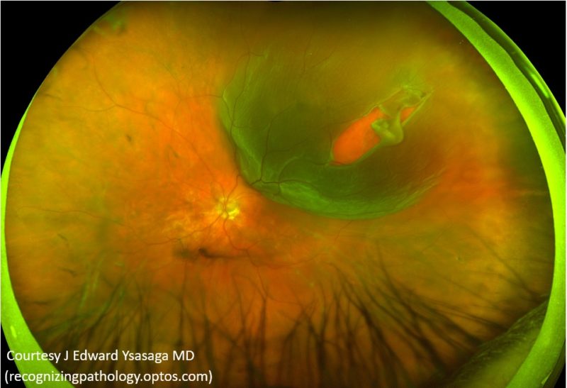
Retinal Detachment
The retinal layer of the eye may sometimes come away from the back wall of the eye as a result of accumulation of fluid. The initiating event is usually a retinal tear as a result of vitreous gel tractional forces within the eye. When the retina is torn, fluid slowly accumulates underneath the retina, peeling it off and causing the detachment. This is perceived by the patient as a black shadow which impairs the field of vision. In early stages of the detachment, central vision is preserved and the patient is only aware of the shadow. As the condition progresses, vision drops and the shadow enlarges in size.
There is no option but to have surgery in order to treat a detached retina. If untreated, the eye will eventually be blind.
The surgical treatment usually involves the removal of the vitreous gel (vitrectomy), lasering or application of freezing treatment around areas where the retina is torn and finally introduction of a gas bubble in the eye to support the retina whilst it heals.
Visual recovery following a retinal detachment surgery is slow and gradual. If the central vision had been involved prior to surgery (macula off detachment), the final vision of the patient is worse than that before the detachment.
In selected cases, the detached retina can be repaired with another type of procedure called scleral buckling. In this case freezing treatment is applied on the outside of the eye. Then a special sponge is sutured on the white part of the eye (sclera). This supports the detached retina which eventually reattaches.
Vitrectomy surgery causes at most times the eye to develop a cataract few months or a year later in which case elective cataract surgery is required.